Testing and treatment of community-acquired pneumonia caused by Streptococcus pneumoniae or Legionella pneumophila
Pneumonia is a leading cause of death in all age groups worldwide.1,2 In affected patients, the air sacs of the lungs fill with fluid or pus, hindering oxygen exchange. There are dozens of reported infectious agents, including bacteria, fungi, and viruses.3,4 Additionally, non-infectious causes such as aspiration of foreign substances or inhalation of chemicals or other irritants have been reported.5,6 Diagnosis and subsequent appropriate treatment can be challenging since symptoms are similar irrespective of the causative agent and include fever, chest pain, cough, abnormal breathing, and rapid heart rate.


Classification of Pneumonia: CAP, HAP, and VAP

Pneumonia is often classified by site of acquisition: community-acquired pneumonia (CAP), hospital-acquired pneumonia (HAP) or ventilator-associated pneumonia (VAP). CAP is the most common and, in the United States, accounts for more than 1.5 million visits annually to emergency departments.7 While symptoms of all classifications are similar, the causative agents are notably different. Antibiotic resistant organisms such as methicillin-resistant Staphylococcus aureus (MRSA) are more commonly observed in HAP and VAP patients,8 while Streptococcus pneumoniae is found in 33-50% of CAP infections in adults and is the most frequently identified organism in specimens taken from patients with CAP.9 Globally, pneumonia is the leading cause of death in children under 5 years old, with an estimated 672,000 deaths annually,10 and the fifth leading cause of death in adults over 70, with an estimated 1.23 million deaths per year.11 If diagnosis of pneumonia caused by S. pneumoniae (pneumococcal pneumonia) is delayed or inaccurate, S. pneumoniae can enter the blood stream, cross the blood-brain barrier, or infect the heart and joints, manifesting as bacteremia, meningitis, pericarditis or arthritis.12

Legionellosis, caused by Legionella pneumophila, is another significant cause of CAP.13 Legionellosis is subdivided into nonpneumonic (Pontiac fever) and pneumonic (Legionnaires’ disease.) Pontiac fever is an acute, self-limiting influenza-like illness, usually lasting 2–5 days with symptoms of fever, chills, headache, malaise, and muscle pain occurring within 72 hours of exposure. Legionnaires’ disease has an incubation period of 2 to 10 days (but up to 19 days has been recorded in some outbreaks) and presents initially with symptoms of fever, loss of appetite, headache, malaise, and lethargy.

Some patients may also experience muscle pain, diarrhea, and confusion. The severity of Legionnaires’ disease ranges from a mild cough to a rapidly fatal pneumonia, with death occurring through progressive pneumonia with respiratory failure and/or shock and multi-organ failure. In 2018, there were nearly 10,000 cases of Legionnaires’ disease in the United States,14 – the most in U.S. history, though this number is likely an underestimate.
Legionnaires’ Disease is underdiagnosed largely due to the difficulty in distinguishing Legionnaires’ disease from other types of pneumonia. The incidence rate is likely 1.8-2.7 times higher than what is reported with a mortality rate of about 10%.15

Microbiology

Streptococcus pneumoniae

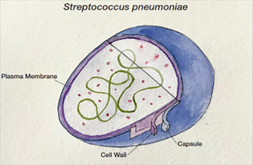
S. pneumoniae bacteria are facultative anaerobic, Gram-positive, lancet-shaped organisms (Figure 1). Typically seen in pairs, or diplococci, they can also occur singularly or in short chains. Most pneumococci are encapsulated, with an outer surface covered by a layer of complex polysaccharides. The polysaccharide capsule is a central virulence factor as well as a major protective antigen of S. pneumoniae and protects the bacterial cell wall from host antibodies and complement in the bloodstream.16,17

Capsular polysaccharides vary between different pneumococci and the organism can be classified by type-specific antisera (SSI Diagnostica, Denmark). By 2020, over one hundred serotypes had been recognized.17 Although most serotypes are known to infect humans, a smaller subset of serotypes cause most illnesses.17 Importantly, antibodies specific to the capsular polysaccharide of a serotype are protective against pneumonia caused by that serotype.17 Despite being protective against opsonization by host antibodies in the blood, S. pneumoniae bacteria rapidly shed the capsule when they encounter innate antimicrobial peptides that are present on the respiratory epithelium.19 The de-capsulated bacteria can then attach and invade the epithelial cell more easily and may also be less subject to anti-capsular immunity. Thus, the role of the capsule as a protector or a discardable cover can vary depending on the locality (bloodstream or mucosal surface).18 The protective role in the bloodstream comes at the price of being a target for antibodies, especially in immunized individuals. The interplay of capsule serotype with infectivity complicates vaccine development and challenges our understanding of how serotype replacements occur after successful vaccine introductions. Serotyping in addition to whole genome sequencing provides critical information for effective vaccine development.

Many serotypes of S. pneumoniae organisms are harbored in the nasopharynx.16 Long term carriage of pneumococci in healthy people is common and numerous studies have found that bacteria can be isolated in up to 90% of healthy individuals.16 This location enables easy airborne transmission of bacteria to uninfected persons. From the nasopharynx, S. pneumoniae can easily infect the middle ear or lungs. Although S. pneumoniae can cause disease directly, the major source of illness is the severe inflammation that the bacteria trigger. The seriousness of pneumococcal disease correlates with the strength of the inflammatory reaction.

Legionella pneumophila

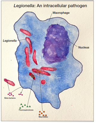
Legionella bacteria are aerobic, unencapsulated, Gram-negative, rod-shaped, intracellular pathogens (Figure 2). Although occurring naturally in small amounts in freshwater environments such as streams and lakes, Legionella poses a health risk when found in water distribution systems of public buildings such as hospitals and hotels. Legionella is also resistant to standard water disinfection procedures and can be found in treated potable water, hot tubs and nearly any water storage device.

These environments allow for Legionella proliferation and the organisms are then transmitted to humans via aerosolization or aspiration of the contaminated water. There are more than 50 Legionella species described in the literature, and 18 of the species have been associated with human infections. Legionella pneumophila is the cause of more than 90% of all cases of Legionnaires’ disease. L. pneumophila is divided into 15 serogroups, with serogroup 1 causing more than 84% of global reported cases of L. pneumophila legionellosis, though this figure is complicated by the fact that some tests only detect serogroup 1 and serogrouping is not routinely performed in many localities.20


Testing

Culture

The gold standard for diagnosis remains traditional culture-based methods, using respiratory samples or blood cultures. The most clear-cut advantage of culture is that positive results provide strong confirmation of organism, which can then be serogrouped or tested for antibiotic resistance. However, obtaining high quality sputum samples is a common limitation,21 such that negative results are typically not informative, particularly in Legionellosis, where less than 50% of patients produce sputum.22-25 Additionally, samples should be collected from the lower airway as upper airway samples can be contaminated.

Blood cultures from patients with pneumonia identify a bacterial pathogen in only 5-16% of cases. This finding is reasonable as not all patients with pneumonia are bacteremic. S. pneumoniae is detected in one third of positive blood cultures.26 Culture results will also be negative for viral pathogens, which appear to cause 30-50% of cases.26 The recent Guidelines of the ATS/IDSA do not recommend blood cultures for adults with nonsevere CAP who are managed as outpatients.21 Prompt initiation of appropriate antibiotic therapy is a goal of treatment for pneumonia illnesses. Culture requires more than 24 hours to identify an organism, or as long as 3-5 days for Legionella, and a further 1-2 days to provide information on its antibiotic sensitivity.27 If antibiotics have been started prior to specimen collection, culture becomes further compromised.

Molecular Panel Assays of Respiratory Pathogens

Multiplex molecular panels target many pathogens, typically from invasive sampling like bronchial lavage, tracheal suction, or sputum samples from lower airways. Inherent broad coverage of pathogens increases the likelihood that an infecting organism will be identified. Panels also incorporate viral pathogens that culture cannot detect and can also include antibiotic resistance genes. Molecular assays are rapid (<2 hours) and highly sensitive. This is useful for infection control in patient settings and outbreak investigations.27 

However, the benefits of comprehensive inclusion and high sensitivity are partially offset because both viral and bacterial pathogens can colonize respiratory sites without causing illness.27 Asymptomatic colonization makes it difficult to clinically assign responsibility and adopt or abandon antibiotic measures for the correct organism, necessitating confirmatory testing.28 Because of the difficulty in obtaining a suitable patient sample and due to the high cost of multiplex panels, these tools are often reserved for the most acutely ill patients.

Urinary Antigen Testing

It may seem counter-intuitive to use a urine sample to detect a respiratory illness, but antigens shed from S. pneumoniae and L. pneumophila can be filtered and concentrated by the kidneys before excretion.29,30 In 1945 Chr. R. Roesgaard wrote a doctoral thesis, “Pneumococcal carbohydrates in urine”, which showed that it was possible to detect pneumococcal antigens in the urine. The first commercial assay became available in 1997. Urinary antigen testing (UAT) detects the antigen in the urine sample, without the need for invasive sampling of the lower respiratory tract site of actual pneumococcal and/or Legionella infection. While L. pneumophila serogroup 1 is responsible for most infections, other serogroups are also pathogenic and not all currently available UAT kits detect non-serogroup 1 cases. It is also important to note that the organism is not itself present in the urine and cannot be cultured from the sample or detected by PCR. Detection of urine antigen is especially useful if antibiotic therapy has already been started.31 Culture of blood or respiratory samples from antibiotic-treated patients will (hopefully) give negative results while antigen is detectable in urine for a longer period of time.29,32 Multiple studies reported that, due to initiation of antibiotic therapy and general culture insufficiencies, culture recovered a pathogen in less than 30% of blood cultures and less than 50% of sputum cultures 33, but a sample produced a positive urine antigen result in as many as 20% of culture-negative patients.29 Such antigen test results should be carefully examined as potential true positives if clinical suspicion and symptoms of pneumonia are present.30,34,35

Urinary antigen testing is non-invasive and offers the simplicity of urine collection and transport. Results can facilitate early diagnosis and help support appropriate treatment, while also potentially providing a pathway to step down from broad-spectrum antibiotic therapies. The simplicity of urine collection and transport, and the accuracy of results supports the early diagnosis and tailored antibiotic treatment of S. pneumoniae and Legionellosis infections. National guidance recommendations have been introduced in some countries to include UAT for both S. pneumoniae and Legionella in addition to X-ray and culture.36,37 A positive test result gives physicians confidence to quickly switch from initial broad-spectrum antibiotic therapy to appropriate, targeted treatment in patients with pneumonia.5,33

Treatment

The mainstay for treatment of bacterial pneumonia and of viral pneumonia in patients at elevated risk for bacterial co-infection remains antibiotics. The most recent Clinical Practice ATS/IDSA Guidelines published in 2019 outline treatment recommendations for community-acquired pneumonia (CAP). These guidelines offer several worthwhile updates because of changes in antibiotic resistance of bacterial pathogens. Viral causes of pneumonia are now widely recognized, and the guidelines note that patients often have viral and bacterial infections simultaneously.21 The recommendations for either type of pneumonia continue to emphasize initial empiric treatment for possible bacterial (co)infection, especially in adult patients with clinical symptoms and radiographic confirmation of pneumonia.

Empiric antibiotics are generally successful for patients with mild CAP, and diagnostic testing to identify a specific pathogen does not usually change treatment or outcomes (Figure 3). Of note, standard empiric therapy for severe CAP now favors beta-lactam/macrolide combinations.21 A new avenue for limiting antibiotic treatment is the use of biomarker tests. Procalcitonin assays can distinguish with about 70% accuracy a viral pneumonia (where antibiotics are not useful and can be discontinued early) from bacterial pneumonia.38 Empiric use of antibiotics, however, is not without risk. A study of the adequacy of initial ceftriaxone antibiotic therapy for CAP found an increased risk of in-hospital death if unnecessarily broad-spectrum antibiotics were used for initial treatment.39 In contrast, inadequate or inappropriate treatment of patients with antibiotic resistant bacteria also carries a risk of mortality.40

While many bacterial pneumonias are treated with penicillin, this antibiotic is not effective against L. pneumophila as it is an intracellular pathogen. Instead, macrolides or quinolones, which can penetrate infected host cells, can be used.41 It is important to identify Legionnaires’ disease and begin treatment as soon as possible, as delay in initiation of antibiotic therapy is a factor associated with higher mortality (Figure 4).

Summary

  • S. pneumoniae and L. pneumophila are serious pathogens that can cause community-acquired pneumonia (CAP) and it is important to differentiate the cause so that appropriate treatment and effective antibiotics can begin quickly.
  • Guidelines of the ATS/IDSA do not recommend blood cultures for adults with nonsevere CAP due to concerns that traditional culture of respiratory or blood samples can fail to identify a bacterial pathogen.
  • Multiplex molecular panels provide broad coverage of pathogens, incorporate viral pathogens that culture cannot detect and also include antibiotic resistance genes. Detection of sub-clinical colonizing pathogens, difficulty obtaining a suitable sample, and cost reduce the utility of these tests.
  • Urinary antigen testing (UAT) detects S. pneumoniae and L. pneumophila antigens shed into urine rather than requiring invasive respiratory samples and can detect the antigens after antibiotics have been started, increasing the detection of illnesses that culture misses to support the early diagnosis and tailored antibiotic treatment of S. pneumoniae infections.
  • Emerging serotypes of S. pneumoniae show reduced susceptibility to both antibiotics and current vaccines. This worrying combination may lead to a rebound in disease.
  • Beta-lactams such as penicillin are not clinically effective when treating Legionella, so antibiotics capable of penetrating host cells, such as macrolides and quinolones, must be used to treat pneumonia caused by L. pneumophila.
  1. References
    1. GBD 2016 Lower Respiratory Infections Collaborators (2018). Estimates of the global, regional, and national morbidity, mortality, and aetiologies of lower respiratory infections in 195 countries, 1990-2016: a systematic analysis for the Global Burden of Disease Study 2016. The Lancet Infectious Diseases, 18, 1191–1210. doi: https://doi.org/10.1016/S1473-3099(18)30310-4
    2. The top 10 causes of death. World Health Organization. https://www.who.int/news-room/fact-sheets/detail/the-top-10-causes-of-death. Published December 9, 2020.
    3. le Roux, D. M., Zar, H. J. Community-acquired pneumonia in children – a changing spectrum of disease. Pediatric Radiology, 2017 Oct;47(11):1392-1398. doi: 10.1007/s00247-017-3827-8.
    4. Musher, D. M., Abers, M.S., Bartlett, J. G. Evolving Understanding of the Causes of Pneumonia in Adults, With Special Attention to the Role of Pneumococcus. Clinical Infectious Diseases, 2017 Oct 30;65(10):1736-1744. doi: 10.1093/cid/cix549.
    5. Marik, P.E. Aspiration pneumonitis and aspiration pneumonia. New England Journal of Medicine. 2001 Mar;344(9):665-671. doi:10.1056/ NEJM200103013440908
    6. Wong J., Magun B.E., Wood L.J. Lung inflammation caused by inhaled toxicants: a review. The International Journal of Chronic Obstructive Pulmonary Disease. 2016 Oct;11:1391-1401. doi: 10.2147/COPD.S106009
    7. Cairns, C., Kang, K. National Hospital Ambulatory Medical Care Survey: 2020 emergency department summary tables. doi: 10.15620/ cdc:121911.
    8. Pickens, C. I., Wunderink, R. G. Methicillin-Resistant Staphylococcus aureus Hospital-Acquired Pneumonia/Ventilator-Associated Pneumonia. Respiratory and Critical Care Medicine, 2022 Apr;43(2):304-309. doi: 10.1055/s-0041-1740583.
    9. Shoar, S., Musher, D.M.. Etiology of community-acquired pneumonia in adults: a systematic review. Pneumonia, 2020 Oct 5;12:11. doi: 10.1186/s41479-020-00074-3
    10. Roser, M., Ortiz-Ospina, E. Causes of death in children under 5. Our World in Data. https://ourworldindata.org/grapher/causes-of-deathin children-under-5. Published in 2019.
    11. Roser, M., Ortiz-Ospina, E. Causes of deaths for people who were 70 years and older. Our World in Data. https://ourworldindata.org/ grapher/causes-of-death-in-70-year-olds. Published in 2019.
    12. Gierke, R., Wodi, A. P., and Kobayashi, M. CDC Pink Book. https://www.cdc.gov/vaccines/pubs/pinkbook/pneumo.html August 2021.
    13. Marchello, C. et al. Prevalence of Atypical Pathogens in Patients With Cough and Community-Acquired Pneumonia: A Meta-Analysis. The Annals of Family Medicine, 2016 Nov;14(6):552-566. doi: 10.1370/afm.1993.
    14. Annual statistics from the National Notifiable Diseases Surveillance System (NNDSS). Centers for Disease Control and Prevention. https://wonder.cdc.gov/nndss/nndss_annual_tables_menu.asp.
    15. Collier, S. A. et al. (2021). Estimate of Burden and Direct Healthcare Cost of Infectious Waterborne Disease in the United States. Emerging Infectious Diseases, 27(1), 140-149. doi: 10.3201/eid2701.190676.
    16. Weiser, J. N., Ferreira, D. M., Paton, J. C. Streptococcus pneumoniae: transmission, colonization and invasion. Nature Reviews Microbiology, 2018 Jun;16(6):355-367. doi: 10.1038/s41579-018-0001-8.
    17. Geno, K. A. et al. Pneumococcal Capsules and Their Types: Past, Present, and Future. Clinical Microbiology Reviews, rev. 2015 Jul;28(3):871-99. doi: 10.1128/CMR.00024-15.
    18. Kietzman, C. C. et al. Dynamic capsule restructuring by the main pneumococcal autolysin LytA in response to the epithelium. Nature Communications, 2016 Feb 29;7:10859. doi: 10.1038/ncomms10859.
    19. Gierke, R. et al. Pneumococcal – vaccine Preventable diseases surveillance manual. Centers for Disease Control and Prevention. http:// www.cdc.gov/vaccines/pubs/surv-manual/chpt11-pneumo.html. Published June 29, 2020.
    20. Yu, V. L. et al. Distribution of Legionella species and serogroups isolated by culture in patients with sporadic community-acquired legionellosis: an international collaborative survey. The Journal of Infectious Diseases, 2002 Jul 1;186(1):127-8. doi: 10.1086/341087.
    21. Metlay, J. P. et al. Diagnosis and Treatment of Adults with Community-acquired Pneumonia. An Official Clinical Practice Guideline of the American Thoracic Society and Infectious Diseases Society of America. American Journal of Respiratory and Critical Care Medicine, 2019 Oct 1;200(7):e45-e67. doi: 10.1164/rccm.201908-1581ST.
    22. Sopena, N. et al. Comparative study of the clinical presentation of Legionella pneumonia and other community-acquired pneumonias. Chest, 1998 May;113(5):1195-200. doi: 10.1378/chest.113.5.1195.
    23. Lieberman, D., et al. Legionella species community-acquired pneumonia. A review of 56 hospitalized adult patients. Chest, 1996 May;109(5):1243-9. doi: 10.1378/chest.109.5.1243.
    24. Woodhead, M. A., Macfarlane, J. T. Legionnaires’ disease: a review of 79 community acquired cases in Nottingham. Thorax, 1986 Aug;41(8):635-40. doi: 10.1136/thx.41.8.635.
    25. Tsai, T. F. et al. Legionnaires’ disease: clinical features of the epidemic in Philadelphia. The Annals of Internal Medicine, 1979 Apr;90(4):509-17. doi: 10.7326/0003-4819-90-4-509.
    26. Klompas, M. Clinical evaluation and diagnostic testing for community-acquired pneumonia in adults. UpToDate. https://www.uptodate. com/contents/clinical-evaluation-and-diagnostic-testing-for-community-acquired-pneumonia-in-adults?topicRef=7027&amp;source= see_link. Updated January 3, 2023.
    27. Poole, S., Clark, T. W. Rapid syndromic molecular testing in pneumonia: The current landscape and future potential. Journal of Infection, 2020 Jan;80(1):1-7. doi: 10.1016/j.jinf.2019.11.021.
    28. Zautner, A. E. et al. More Pathogenicity or Just More Pathogens? – On the Interpretation Problem of Multiple Pathogen Detections with Diagnostic Multiplex Assays. Frontiers in Microbiology, 2017 Jun 29;8:1210. doi: 10.3389/fmicb.2017.01210.
    29. Couturier, M. R., Graf, E. H., Griffin, A. T. Urine antigen tests for the diagnosis of respiratory infections: legionellosis, histoplasmosis, pneumococcal pneumonia. Clinics in Laboratory Medicine, 2014 Jun;34(2):219-36. doi: 10.1016/j.cll.2014.02.002.
    30. Greenfield, A. et al. Impact of Streptococcus pneumoniae Urinary Antigen Testing in Patients with Community-Acquired Pneumonia Admitted Within a Large Academic Health System. Open Forum Infectious Diseases, 2021 Oct 22;9(1):ofab522. doi: 10.1093/ofid/ofab522.
    31. Hasanuzzaman, M. et al. Comparison of Culture, Antigen Test, and Polymerase Chain Reaction for Pneumococcal Detection in Cerebrospinal Fluid of Children. The Journal of Infectious Diseases, 2021 Sep 1;224(12 Suppl 2):S209-S217. doi: 10.1093/infdis/jiab073.
    32. Laijen, W., Snijders, D., Boersma, W. G. Pneumococcal urinary antigen test: diagnostic yield and impact on antibiotic treatment. The Clinical Respiratory Journal, 2017 Nov;11(6):999-1005. doi: 10.1111/crj.12453.
    33. Schimmel, J. J. et al. Pneumococcal Urinary Antigen Testing in United States Hospitals: A Missed Opportunity for Antimicrobial Stewardship. Clinical Infectious Diseases, 2020 Sep 12;71(6):1427-1434. doi: 10.1093/cid/ciz983.
    34. Said, M. A. et al. (2013) Estimating the burden of pneumococcal pneumonia among adults: a systematic review and meta-analysis of diagnostic techniques. PloS ONE, 2013;8(4):e60273. doi: 10.1371/journal.pone.0060273.
    35. Kim, P., Deshpande, A., Rothberg, M. B. Urinary Antigen Testing for Respiratory Infections: Current Perspectives on Utility and Limitations. Infection and Drug Resistance, 2022 Apr 27;15:2219-2228. doi: 10.2147/IDR.S321168.
    36. Overview: Pneumonia in adults: Diagnosis and management: Guidance. NICE. https://www.nice.org.uk/guidance/cg191. Published December 3, 2014.
    37. Mandell, L. A. et al. Infectious Diseases Society of America; American Thoracic Society. Infectious Diseases Society of America/American Thoracic Society consensus guidelines on the management of community-acquired pneumonia in adults. Clinical Infectious Diseases, 2007 Mar 1;44 Suppl 2(Suppl 2):S27-72. doi: 10.1086/511159.
    38. Self, W. H. et al. Procalcitonin as a Marker of Etiology in Adults Hospitalized With Community-Acquired Pneumonia. Clinical Infectious Diseases, 2017 Jul 15;65(2):183-190. doi: 10.1093/cid/cix317.
    39. Van Besien, R. F. et al. Ceftriaxone resistance and adequacy of initial antibiotic therapy in community onset bacterial pneumonia. Medicine, 2022 May 20;101(20):e29159. doi: 10.1097/MD.0000000000029159.
    40. Wongsurakiat, P., Chitwarakorn, N. Severe community-acquired pneumonia in general medical wards: outcomes and impact of initial antibiotic selection. BMC Pulmonary Medicine, 2019 Oct 16;19(1):179. doi: 10.1186/s12890-019-0944-1.
    41. Viasus, D. et al. Legionnaires’ Disease: Update on Diagnosis and Treatment. Infectious Diseases and Therapy, 2022 Jun;11(3):973-986. doi: 10.1007/s40121-022-00635-7.

Additional information

Join our mailing list